
Mouse Secondary Antibodies

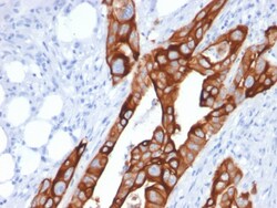
Biotium Cytokeratin 20 (KRT20) (Colorectal Epithelial Marker)(KRT20/1991), 0.2mg/mL
This MAb recognizes an intermediate filament protein of 46 kDa, identified as cytokeratin 20 (KRT20). KRT is abundantly expressed in goblet cells and enterocytes of the gastrointestinal tract. It is a useful marker of pancreatic and colorectal cancer. KRT20 is expressed under normal, hyperplastic and neoplastic conditions. It has been detected in adenocarcinomas of the colon, stomachand biliary tract. Breast carcinomas are generally non-reactive. Primary antibodies are available purified, or with a selection of fluorescent CF Dyes and other labels. CF Dyes offer exceptional brightness and photostability. Note: Conjugates of blue fluorescent dyes like CF405S and CF405M are not recommended for detecting low abundance targets, because blue dyes have lower fluorescence and can give higher non-specific background than other dye colors.

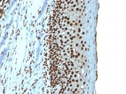
Biotium Histone H1 (Nuclear Marker) (r1415-1), CF488A conjugate, 0.1mg/mL
Eukaryotic histones are basic and water-soluble nuclear proteins that form hetero-octameric nucleosome particles by wrapping 146 base pairs of DNA in a left-handed supealpha-helicalturn sequentially to form chromosomal fiber. Two molecules of each of the four core histones (H2A, H2B, H3, and H4) form the octamer; formed of two H2A-H2B dimers and two H3-H4 dimers, forming two nearly symmetrical halves by tertiary structure. Over 80% of nucleosomes contain the linker Histone H1, derived from an intronless gene that interacts with linker DNA between nucleosomes and mediates compaction into higher order chromatin. Histones are subject to posttranslational modification by enzymes primarily on their N-terminal tails, but also in their globular domains. Such modifications include methylation, citrullination, acetylation, phosphorylation, sumoylation, ubiquitination and ADP-ribosylation.Primary antibodies are available purified, or with a selection of fluorescent CF Dyes and other l
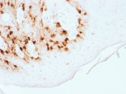
Biotium MART-1 / Melan-A / MLANA (Melanoma Marker) (MLANA/1761R), CF568 conjugate, 0.1mg/mL
This antibody recognizes a protein doublet of 20-22 kDa, identified as MART-1 (Melanoma Antigen Recognized by T cells 1) or Melan-A. MART-1 is a newly identified melanocyte differentiation antigen recognized by autologous cytotoxic T lymphocytes. Seven other melanoma associated antigens recognized by autologous cytotoxic T cells include MAGE-1, MAGE-3, tyrosinase, gp100, gp75, BAGE-1, and GAGE-1. Subcellular fractionation shows that MART-1 is present in melanosomes and endoplasmic reticulum. This MAb labels melanomas and other tumors showing melanocytic differentiation. It is also a useful positive-marker for angiomyolipomas. It does not stain tumor cells of epithelial, lymphoid, glial, or mesenchymal origin.Primary antibodies are available purified, or with a selection of fluorescent CF Dyes and other labels. CF Dyes offer exceptional brightness and photostability. Note: Conjugates of blue fluorescent dyes like CF405S and CF405M are not recommended for detecting low a

Biotium Chromogranin A / CHGA (Neuroendocrine Marker), CF647 conjugate, 0.1mg/mL
Chromogranin A is present in neuroendocrine cells throughout the body, including the neuroendocrine cells of the large and small intestine, adrenal medulla and pancreatic islets. It is an excellent marker for carcinoid tumors, pheochromocytomas, paragangliomas, and other neuroendocrine tumors. Co-expression of chromogranin A and neuron specific enolase (NSE) is common in neuroendocrine neoplasms. Reportedly, co-expression of certain keratins and chromogranin indicates neuroendocrine lineage. The presence of strong anti-chromogranin staining and absence of anti-keratin staining should raise the possibility of paraganglioma. The co-expression of chromogranin and NSE is typical of neuroendocrine neoplasms. Most pituitary adenomas and prolactinomas readily express chromogranin.Primary antibodies are available purified, or with a selection of fluorescent CF Dyes and other labels. CF Dyes offer exceptional brightness and photostability. Note: Conjugates of blue fluorescent dyes
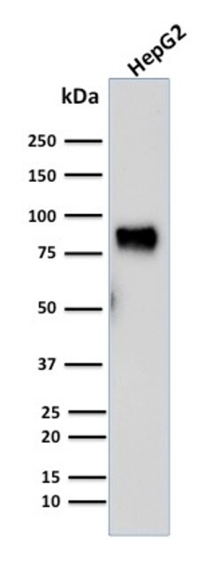
Biotium MDM2(SMP14), CF647 conjugate, 0.1mg/mL
MDM2 is a nuclear phosphoprotein that binds and inhibits transactivation by tumor protein p53. It can promote tumor formation by targeting tumor suppressor proteins, such as p53, for proteasomal degradation. Overexpression of MDM2 can result in excessive inactivation of tumor protein p53, diminishing its tumor suppressor function. This protein also affects the cell cycle, apoptosis, and tumorigenesis through interactions with other proteins, including retinoblastoma 1 and ribosomal protein L5. Overexpression of MDM2 protein is detected in a variety of cancers. Primary antibodies are available purified, or with a selection of fluorescent CF Dyes and other labels. CF Dyes offer exceptional brightness and photostability. Note: Conjugates of blue fluorescent dyes like CF405S and CF405M are not recommended for detecting low abundance targets, because blue dyes have lower fluorescence and can give higher non-specific background than other dye colors.
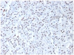
Biotium Wilm's Tumor 1 (WT1) (Wilm's Tumor & Mesothelial Marker), Biotin conjugate, 0.1mg/mL
Recognizes a 47-55 kDa-tumor suppressor protein, identified as Wilm's Tumor (WT1) protein. The antibody reacts with all isoforms of the full-length WT1 and also identifies WT1 lacking exon 2-encoded amino acids, frequently found in subsets of sporadic Wilms tumor and mesothelioma. WT1 protein has been identified in proliferative mesothelial cells, malignant mesothelioma, ovarian carcinoma, gonadoblastoma, nephroblastoma, and desmoplastic small round cell tumor. Lung adenocarcinomas rarely stain positive with this antibody. WT1 protein expression in mesothelial cells has become a reliable marker for the diagnosis of mesotheliomas. Primary antibodies are available purified, or with a selection of fluorescent CF Dyes and other labels. CF Dyes offer exceptional brightness and photostability. Note: Conjugates of blue fluorescent dyes like CF405S and CF405M are not recommended for detecting low abundance targets, because blue dyes have lower fluorescence and can give higher no
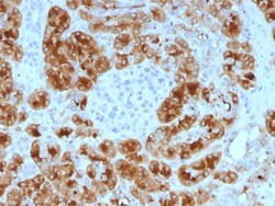
Biotium GP2 (Glycoprotein 2) / ZAP75 (GP2/1805), Biotin conjugate, 0.1mg/mL
GP2 (glycoprotein 2), also known as ZAP75, is a 537 amino acid secreted protein. It is an integral membrane protein that is secreted from intracellular zymogen granules and associates with the plasma membrane via glycosylphosphatidylinositol (GPI) linkage. GP2 is cleaved and then released into the pancreatic duct along with exocrine secretions. GP2 binds pathogens such as enterobacteria, thereby playing an important role in the innate immune response. The C-terminus of this protein is related to the C-terminus of the protein encoded by the neighboring gene, uromodulin (UMOD). GP2 is also expressed on the apical plasma membrane of specialized microfold (M) cells among enterocytes and serves as a transcytotic receptor for mucosal antigens. M cells are considered a promising target for oral vaccination against various infectious diseases.Primary antibodies are available purified, or with a selection of fluorescent CF Dyes and other labels. CF Dyes offer exceptional brightness
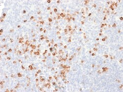
Biotium Human IgG (Immunoglobulin Gamma Heavy Chain) (B-Cell Marker) (rIG266), CF740 conjugate, 0.1mg/mL
Recognizes a protein of 75 kDa, identified as gamma heavy chain of human immunoglobulins. It does not cross-react with alpha (IgA), mu (IgM), epsilon (IgE), or delta (IgD), heavy chains, T-cells, monocytes, granulocytes, or erythrocytes. This MAb is useful in the identification of leukemias, plasmacytomas, and certain non-Hodgkin's lymphomas. The most common feature of these malignancies is the restricted expression of a single heavy chain class. Demonstration of clonality in lymphoid infiltrates indicates that the infiltrate is clonal and therefore malignant. Primary antibodies are available purified, or with a selection of fluorescent CF Dyes and other labels. CF Dyes offer exceptional brightness and photostability. Note: Conjugates of blue fluorescent dyes like CF405S and CF405M are not recommended for detecting low abundance targets, because blue dyes have lower fluorescence and can give higher non-specific background than other dye colors.
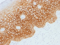
Biotium CD44v4 (Marker of Tumor Metastasis), CF568 conjugate, 0.1mg/mL
This antibody recognizes an epitope encoded by exon v4 on the variant portion of human CD44. The CD44 molecule belongs to a family of cellular adhesion molecules found on a wide range of normal and malignant cells in epithelial, mesothelial and hemopoiesis tissues. CD44 is a single gene with 20 exons, of which 10 are normally expressed to encode the basic CD44 (H-CAM) molecule. The additional 10 exons (v1 to v10) are only expressed by alternative splicing of the nuclear RNA. The expression of specific cell adhesion molecule CD44 splice variants has been reported to be associated with metastasis in certain human malignancies. Primary antibodies are available purified, or with a selection of fluorescent CF Dyes and other labels. CF Dyes offer exceptional brightness and photostability. Note: Conjugates of blue fluorescent dyes like CF405S and CF405M are not recommended for detecting low abundance targets, because blue dyes have lower fluorescence and can give higher non-spe
Biotium Histone H1 (Nuclear Marker) (r1415-1), CF740 conjugate, 0.1mg/mL
Eukaryotic histones are basic and water-soluble nuclear proteins that form hetero-octameric nucleosome particles by wrapping 146 base pairs of DNA in a left-handed supealpha-helicalturn sequentially to form chromosomal fiber. Two molecules of each of the four core histones (H2A, H2B, H3, and H4) form the octamer; formed of two H2A-H2B dimers and two H3-H4 dimers, forming two nearly symmetrical halves by tertiary structure. Over 80% of nucleosomes contain the linker Histone H1, derived from an intronless gene that interacts with linker DNA between nucleosomes and mediates compaction into higher order chromatin. Histones are subject to posttranslational modification by enzymes primarily on their N-terminal tails, but also in their globular domains. Such modifications include methylation, citrullination, acetylation, phosphorylation, sumoylation, ubiquitination and ADP-ribosylation.Primary antibodies are available purified, or with a selection of fluorescent CF Dyes and other l
Biotium AMACR / p504S (Prostate Cancer Marker) (AMACR/1723), CF740 conjugate, 0.1mg/mL
This antibody recognizes a protein of 42 kDa, which is identified as Alpha-methylacyl-CoA Racemase (AMACR), also known as p504S. It is an enzyme that is involved in bile acid biosynthesis and beta-oxidation of branched-chain fatty acids. AMACR is essential in lipid metabolism. It is expressed in cells of premalignant high-grade prostatic intraepithelial neoplasia (HGPIN) and prostate adenocarcinoma. The majority of the carcinoma cells show a distinct granular cytoplasmic staining reaction. AMACR is present at low or undetectable levels in glandular epithelial cells of normal prostate and benign prostatic hyperplasia. A spotty granular cytoplasmic staining is seen in a few cells of the benign glands. AMACR is expressed in normal liver (hepatocytes), kidney (tubular epithelial cells) and gall bladder (epithelial cells). Expression has also been found in lung (bronchial epithelial cells) and colon (colonic surface epithelium). AMACR expression can also be found in hepatocellular
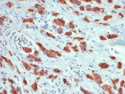
Biotium MUC1 / CA15-3 / EMA / CD227 (Epithelial Marker) (MUC1/1887R), CF488A conjugate, 0.1mg/mL
This MAb reacts with MUC1, a large transmembrane glycoprotein expressed on the ductal surface of normal glandular epithelia. It is used as tracer agent in CA15.3 assays. The extracellular domain of MUC1 largely consists of a highly conserved, O-glycosylated 20 amino acids tandem repeat which can occur 30-100 times per molecule depending on the length of the allele involved. In the vast majority of human carcinomas this protein is up-regulated and poorly glycosylated and appears on the cell surface in a non-polarized fashion. The dominant epitope of this MAb involves both amino acids as well as sugar moieties. Its epitope is destroyed by desialylation i.e. treatment with Neuraminidase.Primary antibodies are available purified, or with a selection of fluorescent CF Dyes and other labels. CF Dyes offer exceptional brightness and photostability. Note: Conjugates of blue fluorescent dyes like CF405S and CF405M are not recommended for detecting low abundance targets, because b
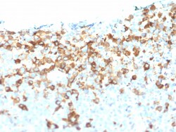
Biotium NGF-Receptor (p75) / CD271 (Soft Tissue Tumor Marker) (NGFR/1964), CF568 conjugate, 0.1mg/mL
This antibody recognizes a glycoprotein of 75 kDa, identified as low affinity Nerve Growth Factor (NGF) Receptor (p75NGFR) or Neurotrophin Receptor (p75NTR). NGFR is expressed in various neural crest cells and their tumors such as melanocytes, melanomas, neuroblastomas, pheochromocytomas and neurofibromas. Reportedly, anti-NGFR is a reliable marker for desmoplastic and neurotropic melanomas. NGFR is expressed in mature non-neural cells such as perivascular cells, dental pulp cells, lymphoidal follicular dendritic cells, basal epithelium of oral mucosa and hair follicles, prostate basal cells, and myoepithelial cells. Anti-NGFR stains the myoepithelial cells of breast ducts and intra-lobular fibroblasts of breast ducts.Primary antibodies are available purified, or with a selection of fluorescent CF Dyes and other labels. CF Dyes offer exceptional brightness and photostability. Note: Conjugates of blue fluorescent dyes like CF405S and CF405M are not recommended for det
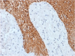
Biotium Cytokeratin, Multi (Epithelial Marker)(KRT/1877R), 0.2mg/mL
Twenty human keratins are resolved with two-dimensional gel electrophoresis into acidic (pI 6. 0) subfamilies. This antibody recognizes acidic (Type I or LMW) and basic (Type II or HMW) cytokeratins, including 59 kDa (CK4); 58 kDa (CK5); 56 kDa (CK6); 52 kDa (CK8); 56. 5 kDa (CK10); 53 kDa (CK13) and 45 kDa (CK18). This is a broad-spectrum antibody, which has been reported to differentiate epithelial tumors from non-epithelial tumors. Many studies have shown the usefulness of keratins as markers in cancer research and tumor diagnosis. Primary antibodies are available purified, or with a selection of fluorescent CF Dyes and other labels. CF Dyes offer exceptional brightness and photostability. Note: Conjugates of blue fluorescent dyes like CF405S and CF405M are not recommended for detecting low abundance targets, because blue dyes have lower fluorescence and can give higher non-specific background than other dye colors.
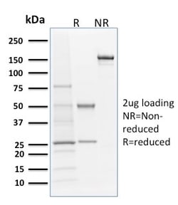
Biotium Langerin / CD207 (Marker of Langerhans Cells)(LGRN/1821), 0.2mg/mL
Langerhans cells (LCs) are a subset of immature dendritic cells (DCs) that specifically localize in the epidermis and other mucosal epithelia. Epidermal LCs possess strong immuno-stimulatory capacity and play a central role in the initiation and regulation of immune responses. Langerin (CD207) is a Ca2 -dependent, C-type lectin domain containing, type II transmembrane protein that induces epidermal LCs to differentiate into Birbeck granules (BG). BGs are organelles with superimposing and zippering membranes that influence proper class I type antigen presentation to the circulating T cells. Human spleen, lymph node, thymus, liver, lung and heart express Langerin protein. Primary antibodies are available purified, or with a selection of fluorescent CF Dyes and other labels. CF Dyes offer exceptional brightness and photostability. Note: Conjugates of blue fluorescent dyes like CF405S and CF405M are not recommended for detecting low abundance targets, because blue dyes have